Module 5: Introduction to Microbiology Stains

Module 5.1: Cytological Stains

Introduction to stains

There are myriad of stains that are available in veterinary medicine for the diagnosis of various diseases. These stains can be divided into two broad categories based on specific goals. These include those that are used for cytology or evaluating cellular morphology and microbial stains.

Cytological stains: Romanowsky type stain

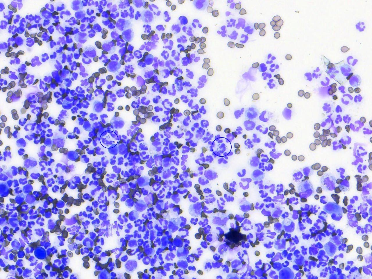
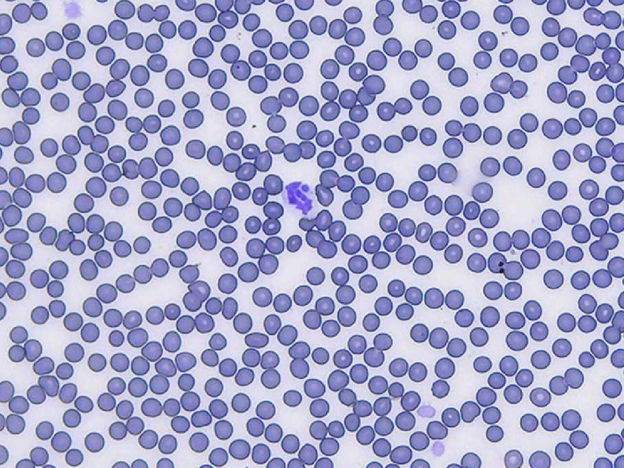
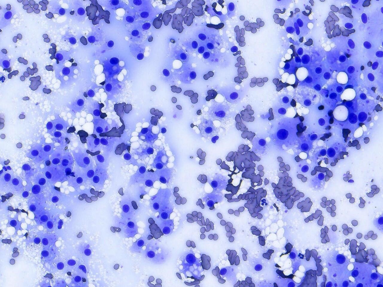
In veterinary medicine, the most common cytological stains are Romanowsky-type stains, such as Diff Quik or Wright-Giemsa stains. The goal of cytological staining is to highlight cellular morphology, specifically nuclear and cytoplasmic details. While these stains do stain common microbes and can be especially useful for identifying protozoal, microfilaria, and fungal agents, these stains are particularly useful in characterizing host structures, such as inflammatory cells (neutrophils and macrophages), neoplastic cells, or resident cellular components. All bacteria, regardless if Gram-positive or negative stain blue or deep purple using Romanowsky types stains.

Fine-needle aspirates from an abscess in a dog. In this image, you can see that there is marked suppurative inflammation characterized by many degenerate neutrophils and low numbers of macrophages. Degenerate neutrophils containing phagocytized bacteria are circled. 100x objective
Fine-needle aspirates from an abscess in a dog. In this image, you can see that there is marked suppurative inflammation characterized by many degenerate neutrophils and low numbers of macrophages. Degenerate neutrophils containing phagocytized bacteria are circled. 100x objective, Diff Quik.


Canine blood smear with a single segmented neutrophil that contains a single Anaplasma phagocytophilum morula. Tee morula is the blue, circular structure on the northern end of the cell. You can appreciate that the bacteria in the abscess and the bacterial morulae in this smear are the same blue color. 100x objective
Canine blood smear with a single segmented neutrophil that contains a single Anaplasma phagocytophilum morula. The morula is the blue, circular structure on the northern end of the cell. You can appreciate that the bacteria in the abscess and the bacterial morulae in this smear are the same blue color. 100x objective, Diff Quik.


Liver aspirates from a dog with well-differentiated hepatocellular carcinoma. You can see that the neoplastic hepatocyte nuclei are round, 2x the diameter of an RBC and placed in the center of the cells. The cytoplasm is the pale blue color that surrounds the nuclei. Sometimes the cytoplasm has variably sized lipid droplets within the cytoplasm. 50x objective
Liver aspirates from a dog with well-differentiated hepatocellular carcinoma. You can see that the neoplastic hepatocyte nuclei are round, 2x the diameter of an RBC, and placed in the center of the cells. The cytoplasm is the pale blue color that surrounds the nuclei. Sometimes the cytoplasm has variably sized lipid droplets within the cytoplasm. 50x objective, Diff Quik

Procedure:

In the laboratory, you will be given step-by-step instructions on how to perform the Diff Quik procedure. Here is a video that also explains the steps.


Knowledge check

License

Icon for the Creative Commons Attribution-NonCommercial 4.0 International License

Clinical Veterinary Diagnostic Laboratory Copyright © 2021 by Erin Burton is licensed under a Creative Commons Attribution-NonCommercial 4.0 International License, except where otherwise noted.

Share This Book