
Module 7: Hematogenous Infectious Disease
Module 7.1: Blood Smear Evaluation for Infectious Disease
Blood smear evaluation for infectious disease
When evaluating a blood smear for infectious agents, you follow the same flow as you would for acute leukemia or immune-mediated hemolytic anemia. As a reminder, the blood smear is comprised of 3 different “zones”; the feathered edge, monolayer, and the body. The smear exam begins with a “flyover” at low magnification (10x objective) of the entire slide. While this quick evaluation may seem like a waste of 30 seconds, it is by far the most important step of any blood film evaluation. At low magnification, you can identify large cells, confirm a leukocytosis described by your hematology analyzer, and infectious components that might otherwise be missed. Classic infectious examples of these are Dirofilaria immitus or Acanthocheilonema reconditum, both microfilariae found in the blood of dogs in the United States and seen best at low magnification.
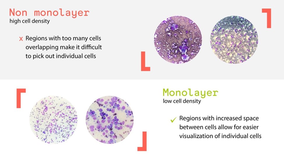
Once scanning the entire slide using the 10x objective, you will move on to the 40x objective to evaluate the WBC’s. WBC’s that contain phagocytized bacteria (bacteremia), rickettsia organisms (Anaplasma or Ehrlichia sp.), fungi (Histoplasma sp.), or protozoal organisms (Leukocytozoon) are easiest found in the monolayer (See figure below on how to locate the monolayer) and the feathered edge. The organisms result in the WBC’s being slightly heavier and they end up migrating towards the feathered edge. Additionally, extracellular organisms that are >10 μm are evaluated at this level. Examples of an extracellular organism seen best at high power include Trypanosoma sp. (T. cruzi, T. congolense, T. vivax, T. brucei subsp. brucei, and T. simiae)
Last, the blood smear is evaluated within the monolayer at the 100x objective (oil immersion). At high magnification, organisms that are <3 μm (most neutrophils are 15 μm) are easiest to visualize. Examples of these organisms include; Babesia sp., Cytauxzoon felis, Mycoplasma sp., and several others.
Key Takeaways
- Step 1: Evaluate the entire slide at low power using your 10x objective lens
- Step 2: Evaluate the WBC in the monolayer high power using you 40x objective lens (no oil)
- Step 3: Evaluate the RBCs and platelets at high power using your 100x objective lens (immersion oil required)
