
Module 10: Veterinary Ectoparasites
Mites
Mite Learning Objectives
- Describe the life cycle stages of mites.
- What are the primary clinical signs of mite infestations?
- How can visual inspection and microscopic examination be used to diagnose mite infestations?
Mites are small arachnids that infest various parts of their hosts, causing a range of dermatological and systemic issues. This section will provide an overview of the biology, identification, common types, and management of the mites.
Types of mites
Mites are categorized into several families based on their morphology, host preference, and the type of pathology they cause. The primary types of mites that affect animals and humans include (Table 10.7):
- Sarcoptidae (Burrowing Mites):
- Examples: Sarcoptes scabiei (scabies mite), Notoedres cati (feline scabies mite).
- Psoroptidae (Non-Burrowing Mites):
- Examples: Psoroptes ovis (sheep scab mite), Chorioptes bovis (cattle chorioptic mange mite).
- Demodicidae (Follicle Mites):
- Examples: Demodex canis (dog follicle mite), Demodex folliculorum (human follicle mite).
- Cheyletiellidae (Fur Mites):
- Examples: Cheyletiella yasguri (dog fur mite), Cheyletiella blakei (cat fur mite).
- Otodectes (Ear Mites):
- Example: Otodectes cynotis (common ear mite in cats and dogs).
Family |
Location in host integument |
Common Species |
|---|---|---|
Sarcoptidae |
Burrow into the skin | Sarcoptes scabiei |
Psoroptidae |
Reside on skin | Octodectes cynotis, Chorioptes sp., Psoroptes sp. |
Cheyletidae |
Reside on skin | Cheyletiella sp. |
Demodecidae |
Reside in hair follicles | Demodex sp. |
Similar to ticks, these arachnids undergo gradual metamorphosis and have 4 pairs (8 legs). Different than adult ticks, the majority of mites are microscopic and difficult to see with the naked eye. Therefore, the microscopic evaluation of skin scrapings (or sometimes scotch tape preps) is the first diagnostic test we reach for with suspected mite infestation.
Life Cycle
The life cycle of mites typically includes the following stages: egg, larva, nymph, and adult. The duration and specifics of each stage can vary among different mite species.
- Egg Stage:
- Female mites lay eggs on the host or in the environment.
- Eggs hatch into larvae within a few days.
- Larva Stage:
- Larvae have three pairs of legs and are usually very small.
- They feed and then molt into nymphs.
- Nymph Stage:
- Nymphs have four pairs of legs and resemble smaller adults.
- They go through one or more molts before reaching adulthood.
- Adult Stage:
- Adults are sexually mature and continue the cycle by mating and laying eggs.
Common clinical signs and pathogenesis
Mite infestations can lead to a variety of clinical signs, depending on the mite species and the severity of the infestation.
- Pruritus (Itching):
- Caused by mite feeding and movement on or within the skin.
- Leads to scratching, biting, and rubbing behaviors.
- Alopecia (Hair Loss):
- Resulting from excessive grooming, self-trauma or in some cases the mite itself.
- Dermatitis:
- Skin becomes red, inflamed, and scabby.
- Secondary bacterial infections may occur due to skin damage.
- Hyperkeratosis and Lichenification:
- Thickening and hardening of the skin due to chronic irritation.
- Specific Clinical Signs for Ear Mites (Otodectes cynotis) in dogs and cats:
- “Coffee ground” appearance of ear discharge.
- Waxy, dark brown, dry parchment-like material in the ears.
- Scratching of ears and scabbed outer pinnae.
- Ear canal hyperkeratosis and hyperplasia.
Diagnosis
- Gross Visual Inspection:
- Observing mites or lesions on the skin, hair, or in the ears.
- Common sites include the head, ears, neck, and limbs.
- Skin Scraping:
- A small area of skin is scraped to collect mites for microscopic examination.
- Collect from the leading edge of the lesion
- Effective for diagnosing burrowing and follicle mites
- Often first diagnostic test chosen for mites
- Microscopic Examination:
- Identifying mites, eggs, larvae, and nymphs under a microscope.
- Essential for accurate identification.
- Otoscope Examination:
- Used specifically for diagnosing ear mites.
- Mites can be observed directly within the ear canal.
- Acetate Tape Impression:
- Tape is applied to the skin to collect surface mites for microscopic examination.
- Not recommended for burrowing or follicular mites
Microscopic Mite Identification
In the laboratory, we will be identifying mites based on the adult forms. An important first step in mite identification is narrowing the differential based on lesion distribution. Mites tend to infect certain aspects of the dermis, epidermis, and adnexal structures (Table 10.7). Similar to ticks, these arachnids undergo gradual metamorphosis, and adults and nymphs have 4 pairs (8 legs). Different than adult ticks, the majority of mites are microscopic and difficult to see with the naked eye. Therefore, the microscopic evaluation of skin scrapings (or sometimes scotch tape preps) is the first diagnostic test we reach for with suspected mite infestation.
Below is Table 10.8 of the common mites of veterinary importance. Salient morphological features are listed below and will help guide you through the identification of the mites in the laboratory exercises.
Species of Mite |
Family of Mite |
Location on host |
Transmission |
Host |
Identifying characteristics |
|---|---|---|---|---|---|
Sarcoptes scabiei (Itch mite) |
Sarcoptidae | Burrow into the epidermis | Direct contact | Mammals | Globose in shape, 3rd and 4th pairs of legs are short |
Otodectes cynotis (ear mites) |
Psoroptidae | Reside on the skin or within the ear | Direct contact | Cat and dog | Males have two suckers ventrally, females have long hairs attached to 3rd and 4th pair of legs |
Cheyletiella spp. (walking dander) |
Cheyletidae | Reside on skin | Direct contact- species specific | Cat, dog, rabbit, humans | Large claws |
Demodex spp. |
Demodecidae | On hair follicle | Skin to skin contact- normal inhabitant | Mammals | Cigar-shaped, 8 legs toward the head |
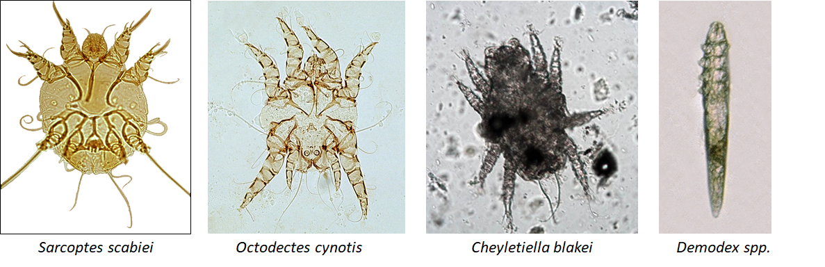 Examples of Common Mite Species- Note that in this image Otodectes is spelled incorrectly
Common mites in veterinary medicine
Sarcoptes Scabiei (Sarcoptic Mange Mite)
- Hosts: Mammals
- Symptoms: Intense itching, rash, hair loss, and skin crusting.
- Diagnosis: Skin scraping and microscopic examination.
Demodex spp. (Demodectic Mange Mite)
- Hosts: All mammals
- Symptoms: Hair loss, redness, and in severe cases, skin infections.
- Diagnosis: Deep skin scraping and microscopic examination.
- Hosts: Dogs, cats, and ferrets
- Symptoms: Ear irritation, scratching, dark discharge, and secondary infections.
- Diagnosis: Otoscopic examination and microscopic examination of ear discharge.
Psoroptes spp. (Psoroptic Mange Mite)
- Hosts: Livestock such as sheep, cattle, and horses. Also the cause of ear mites in rabbits.
- Symptoms: Severe itching, hair loss, and crusting lesions.
- Diagnosis: Skin scraping and microscopic examination.
Cheyletiella spp. (Walking Dandruff Mite)
- Hosts: Dogs, cats, rabbits, humans
- Symptoms: Dandruff, itching, and hair loss.
- Diagnosis: Tape test and microscopic examination.
Summary
Mites are significant ectoparasites that can cause severe dermatological and systemic issues in a wide range of hosts. Understanding the life cycles, clinical signs, and effective control measures for different mite species is crucial for managing infestations and ensuring the health and well-being of affected animals and humans. Regular monitoring, prompt treatment, and integrated pest management strategies are essential for successful control and prevention of mite infestations.
Knowledge check
This is a type of development in which the immature stage (nymph) is a smaller version of the adult.
