
Module 13: Intro to Dermatophytes
Module 13.3: Three Most Common Veterinary Dermatophytes Observed on DTM
The three most common veterinary dermatophytes observed on DTM
In the laboratory you will be asked to identify the 3 most common dermatophytes commonly cultured on DTM; Microsporum canis, Microsporum gypseum, and a Trichophyton sp. Below are descriptions and images of those fungi.
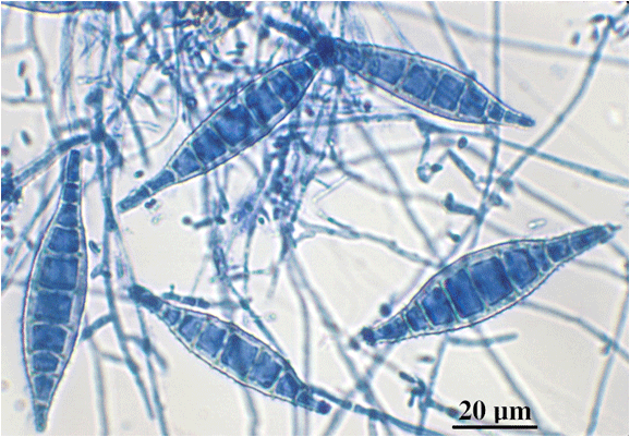
Microsporum canis
The macroconidia are multicellular (5-15 cells), spindle-shaped with rough, thick-walled. The macroconidia have a terminal knob. They produce fewer macroconidia than M. gypseum.
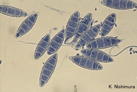
Microsporum gypseum
The macroconidia are numerous, multicellular (4-6 cells), symmetrical, ellipsoidal, with thin walls. The ends of the macroconidia differ in shape. The distal end is rounded where the proximal end can be truncated from where it attached to hyphae.
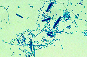
Trichophyton sp.
The microconidia are single-celled, spherical in shape, and numerous. The macroconidia are cigar-shaped, multicellular (2-5 cells), thin-walled, and are rarely seen. These can be differentiated from M. canis as they have 6 compartments formed by the septae.
Knowledge check
Key Takeaways
- Dermatophytes are common cutaneous fungal pathogens in all species
- Microsporum and Trichophyton are the 2 most common genera in veterinary medicine
- On DTM, the dermatophyte fungal colonies are buff in color and accompany a color change to red on the agar
- Saphyrophytic or contaminant fungi will grow on DTM, but do not routinely cause a color change and the colonies are not buff
- Microscopic evaluation of the colonies is an important step in the diagnosis and may influence the control steps based on the species of dermatophyte observed (i.e. geophilic vs. zoophilic)
